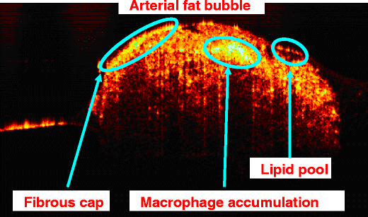
In addition, the subsequent retinal layer segmentation also shows that the proposed method makes the automatic retinal layer segmentation more accurate. Experimental results show that the proposed method performs better than other methods. The proposed method is evaluated through the mean square error, peak signal to noise ratio and the mean structure similarity index using high quality line-scan images as reference. Then low rank matrix completion using bilateral random projection is utilized to iteratively estimate the noise and recover the underlying clean A -scan. In the method, the neighboring A -scans are aligned/registered to the A -scan to be reconstructed and form a matrix together. Based on the assumption that neighboring A -scans are highly similar in the retina, the method reconstructs each A -scan from its neighboring scans. The proposed method models each A -scan as the sum of underlying clean A -scan and noise. In this paper, we propose a new method for speckle reduction in 3D OCT. Therefore, it is unpractical in 3D scan as it requires a much longer data acquisition time. However, it leads to an increase of the data acquisition time.

Overlapping scan is often used for speckle reduction in a 2D line-scan. A raw OCT image/volume usually has very poor image quality due to speckle noise, which often obscures the retinal structures. It has been widely used for retinal imaging in ophthalmology. Optical coherence tomography (OCT) is a micrometer-scale, cross-sectional imaging modality for biological tissue.


 0 kommentar(er)
